Phase-Contrast Imaging for the study and characterization of cultural heritage materials: application of an innovating laboratory-based setup to investigate artifacts from museums and other institutions
Over the last few decades, X-ray imaging has improved significantly as a high-resolution and non-invasive tool for inspecting the internal structure of different kinds of materials. The conventional absorption contrast method is particularly beneficial for characterizing medium density samples or distinguishing materials
with different attenuation powers but leads to poor image quality when the sample is weakly absorbing, typically in materials composed of light elements. On the contrary, Phase-Contrast (PC) X-ray Imaging is an effective technique for detecting low contrast details in weakly absorbing samples, as it provides high-quality
information on micro-structural features. This open interesting prospects in the cultural heritage field. A coherent X-ray beam is the ideal tool to develop phase-sensitive X-ray imaging, and some techniques have already been implemented at Synchrotron Radiation (SR) facilities. Multiple studies already demonstrated the
higher performance of PC-Computed Tomography (CT) with synchrotron radiation, with respect to conventional tomography [Stevenson, 2003; Mocella, 2015; Romell, 2018]
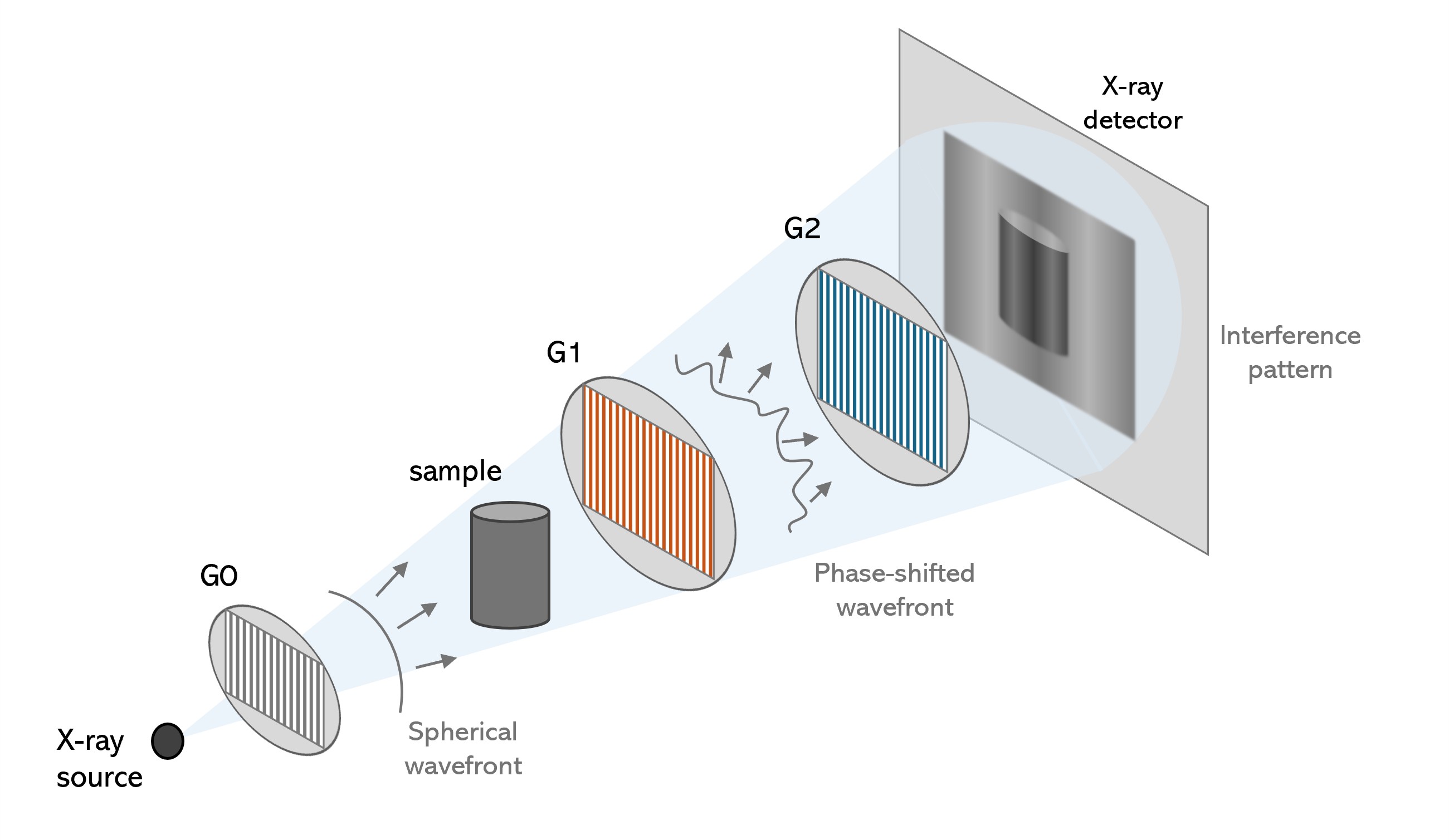
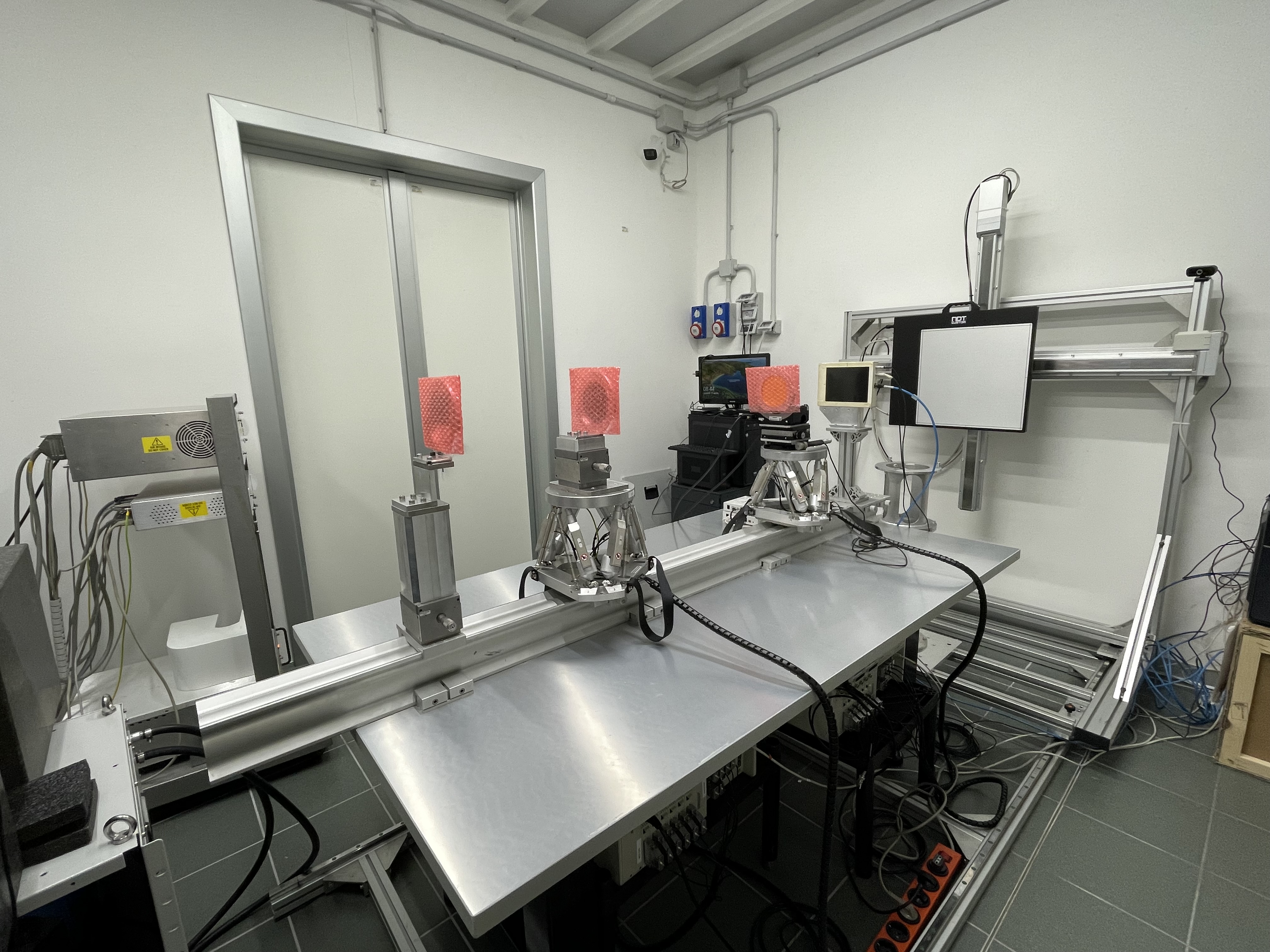
This PhD research is part of the PITCH project funded by MUR in the framework of PRIN2022. The University of Torino together with the National Institute of Nuclear Physics INFN-TO and other institutions have been developing an X-ray Grating Interferometry (GI) laboratory setup based on a Metal-jet X-ray source [Hemberg, 2003].
The research aims are 1) validating the new Phase-Contrast X-ray Imaging setup, 2) benchmarking the image quality obtained with PITCH against synchrotron PC-imaging (PCI) and comparing the obtained results with complementary neutron imaging, 3) optimizing the setup to analyze different cultural heritage (CH) materials to
characterize them and understand related issues, and 4) characterizing samples relevant to cultural heritage preservation projects, prioritizing requests from national research groups and institutions.
Thanks to the participation of a sub-unit of the CCR La Venaria Reale in the project, the creation of CH mock-ups will be carried out together with a selection of relevant materials and original fragments e.g. paper, parchment, cartonnage, textile yarns, wood, layered paintings, metallic yarns, leather, mummified remains.
Different mockup geometries will be tested as well. These analyses will be carried out to characterize the materials and to understand related issues e.g. degradation byproducts, porosity, state of conservation, production techniques.
The last step of the research will be the characterization and analysis with the new system of real artifacts. Different museums will be involved to provide artifacts selected according to their interests and availability.
The development of a lab-based PC-CT system will significantly extend the opportunity to investigate, by a non-invasive approach, both materials and artifacts from the archaeological and the cultural heritage domain. With the new system, laboratory analysis of artworks and archaeological finds with PC-CT,
now confined to a few synchrotron facilities, will be possible, more accessible and feasible. The output of the project would help archaeologists, anthropologists, art historians and restorers in better understanding the realization techniques, the materials and their state of conservation.
References[Stevenson, 2003] A. W. Stevenson et al., "Phase-contrast X-ray imaging with synchrotron radiation for materials science applications", Nucl. Instrum. Methods Phys. Res. Sect. B Beam Interact. Mater. At., vol. 199, pp. 427-435, gen. 2003, doi: 10.1016/S0168-583X(02)01557-4.[Mocella, 2015] V. Mocella et al., "Revealing letters in rolled Herculaneum papyri by X-ray phase-contrast imaging." Nat Commun 6, 5895, 2015.[Romell, 2018] J. Romell et al., "Soft-Tissue Imaging in a Human Mummy: Propagation-based Phase-Contrast CT." Radiology, 289(3):670-676, dec. 2018, doi: 10.1148/radiol.2018180945.[Hemberg, 2003] O. Hemberg et al., "Liquid-metal-jet anode electron-impact x-ray source", Appl. Phys. Lett., vol. 83, fasc. 7, pp. 1483-1485, ago. 2003, doi: 10.1063/1.1602157.[Patera, 2022] A. Patera et al., "X-ray grating interferometry design for the 4D GRAPH-X system", J. Phys. Appl. Phys., vol. 55, fasc. 4, p. 045103, gen. 2022, doi: 10.1088/1361-6463/ac2fd6.